PRRS – past, present and future
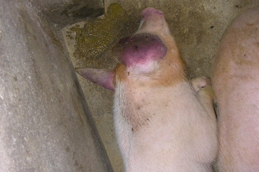
It is too simple to speak of ‘just’ Porcine Reproductive and Respiratory Syndrome (PRRS). History teaches us that the virus changes and various types and subtypes have their own specific characteristics. How can this knowledge help to beat the virus?
By Dr Hans Nauwynck, Laboratory of Virology, Faculty of Veterinary Medicine, Ghent University, Merelbeke, Belgium
A new disease characterised by reproductive and respiratory problems emerged in Northern America and Western Europe in the late 1980s, early 1990s. It was caused by a porcine arterivirus, which based on the symptoms was called Porcine Reproductive and Respiratory Syndrome virus (PRRSv).
On the two continents, two clearly different genetic/ antigenic viruses were circulating: An American type and a European type. Based on serological examinations, it was shown that the American type circulated already earlier in Northern America and by genetic analysis, more genetic variation was detected for the American type in Northern America than for the European type in Western Europe. This latter finding could be attributed to the earlier circulation and/or multiple introductions of the virus in the American pig population.
Germany
Western Europe was confronted with a single introduction, starting in Western Germany. As a consequence, the early European type strains were genetically closely related. In the early 1990s, the American type was proven to be more pathogenic than the European type. Indeed, whereas both virus types had the same power to give reproductive problems upon infection during late gestation, the American type was giving more general clinical signs (fever, anorexia) and respiratory problems than the European type. Only in combinations with other pathogens/ toxins the European type was able to induce overt general and respiratory clinical signs.
By recombination and genetic drift, American strains evolved fast, giving rise to new strains that were even more virulent and extremely difficult to control by commercial vaccines (atypical PRRSv, Sow Abortion and Mortality Syndrome (SAMS)). In 2006, extremely aggressive variants of the American type appeared in China, which are now damaging the whole Asian pig population (long lasting high fever, respiratory and reproductive problems, high mortality) and which represent a real threat to other continents.
Eastern Europe
Research by Dr Uladzimir Karniychuk in 2009 showed that in Eastern Europe, a surprisingly large variation was found for the European type isolates leading to the identification of new subtypes (2, 3 and 4) that were quite different from subtype 1. At present, it is hypothesised that the European type was circulating in Eastern Europe a long time before the entrance of subtype 1 in Western Europe. When the whole group of Western and Eastern European PRRSv strains are considered, a genetic variation of the European type was found that was even larger than the one found with the American type in the US, leading to a very complex genetic world picture of PRRSv.
Eastern European PRRSv strains of subtype 2 (prototype Bor) and 3 (prototype Lena) have been shown to be more virulent than the Western European strains (subtype 1, also known as prototype Lelystad). The European type strain Lena is even as virulent and pathogenic as the high fever disease of the American type in Asia. Over the years in Western Europe, the European type remained mainly linked with reproductive problems. Fever and respiratory problems were absent upon experimental single inoculations. However, starting from mid-2013, PRRSv is responsible for flu-like problems in nurseries in Belgium and most probably also neighbouring countries. Upon experimental inoculation with one of these isolates, called Flanders 13, fever and respiratory problems were reproduced. Genetically, this virus is quite different from other circulating PRRS viruses.
Differentiated macrophages
The pathogenesis of PRRSv is fully determined by differentiated macrophages, which are cells that form part of the immunity system within a pig’s body. During the past 20 years, the European-type strains of subtype 1 (prototype Lelystad) replicated very similarly in a pig. Targets are differentiated macrophages that are carrying a so-called ‘sialoadhesin receptor’. These cells can easily be found in tonsils and lungs, lymph nodes, spleen, maternal endometrium and foetal placenta and at lower levels in all other tissues of the pig. Because the virus does not replicate well in the upper respiratory tract, the virus is difficult to isolate from nasal swabs and the virus does not spread fast between pigs.
The European type strains of subtype 3 (prototype Lena) differ from LV-like strains because they are able to infect a new subset of differentiated macrophages that do not possess the sialoadhesin receptor. An additional receptor is most probably responsible for this.
By experiments in nasal mucosa explants it was found that the additional subset is present at high concentrations in and under the respiratory epithelial cells of the upper respiratory tract, allowing a much stronger replication in respiratory tissues (up to 10-100x higher) and giving rise to a strong viral shedding and a fulminant viraemia (100x higher).
Based on the localisation of this new subset of susceptible macrophages, it is hypothesised that they represent nasal macrophages. These cells are forming a dense network and are taking care of the first line of defense against pathogens. Destroying both nasal and alveolar lung macrophages is most probably the reason why the European type Lena has been associated with secondary bacterial infections and sepsis.
New Flanders strains
The new Flanders 13-like strains are the European type subtype 1 strains that are also evolving in the same direction as Lena. By using the nasal mucosa explants, this virus also replicated in non-sialoadhesin positive macrophages. The virus titres in nasal secretions from the European type infected animals are in line with the replication of the virus in nasal mucosa explants. Whereas it is difficult to detect LV-like the European type subtype 1 in nasal secretions, it is very easy to do so with the more virulent the European type subtype 3 and Flanders 13-like strains. Transmission experiments with these different strains are ongoing in order to find out if the power of the virus to replicate in the nasal macrophages may be related to its aerogenic spread. The increase of macrophage subsets that are infected with the European type in time is a dangerous evolution. A ten- to hundredfold increase of replication gives rise to viral mutants with the same magnitude.
Mutagenesis
The process of changing genetic information of an organism – or mutagenesis – is helping the virus to escape from immunity. This is increasing the risk that highly virulent strains is bringing Europe, in a very dangerous situation. In addition, because of the close relationship of the porcine receptors sialoadhesin and CD163 with their human homologs, one should consider the risk of a species jump to humans.
With these interesting findings with the European type strains, researchers at Ghent University have recently performed experiments with the American type strains in nasal mucosa explants. It was found that both old and more recent American type strains from the US easily replicate to high levels in both sialoadhesin positive and sialoadhesin negative macrophages in the nasal mucosa.
This finding is explaining several things. The American type behaved differently from the European type from the very beginning; it replicated in more subsets of macrophages than the old subtype 1 the European type LV-like strains. Its high replication in the upper respiratory tract differs from the low level of replication of the old subtype 1 the European type strains. This also explains why it was and still is very easy for the American type to spread via airborne transmission. In addition, the higher number of macrophage subsets that are infected with the American type explains why it is more virulent/pathogenic than the old subtype 1 the European type strains and give more rise to secondary infections. It also explains why the American type is able to replicate in sialoadhesin-negative pigs.
Immunity
PRRSv is a difficult target for the immunity. Several branches of the immunity have been shown not to be induced or not to be functional. Low levels of interferons are induced. Antibodies are raised starting from eight days post-infection, but it takes several weeks before a weak neutralisation can be demonstrated. Natural killer cells and cytotoxic T-lymphocytes are not sufficiently effective. Only neutralising antibodies, which appear after one month and at low levels together with a not yet identified porcine killer cell are the two branches that still can do the job and should be activated by vaccination. The drift of the virus makes it a moving target and complicates the whole vaccination strategy.
In the near future, it is important to have access to vaccines that are adaptable, enclosing strains that are closely related to the strains circulating in the field. The technologies for making effective adaptable inactivated and attenuated vaccines became available and should be urgently implemented in the field.
The ultimate dream is the development of an adaptable marker vector vaccine (cassette system) that induces a local immunity in the respiratory tract. As long as PRRSv does not persist such as its sister-arterivirus, lactate dehydrogenase elevating virus (LDv), we should keep on going with the investment to develop vaccines.
In conclusion, it is extremely important to better control PRRS in the near future and not to wait till a complete catastrophe occurs.
There is an urgent need for adaptable inactivated and attenuated marker vaccines and improved biosafety measures in order to fully control PRRSv circulation. Pig producers, PRRS researchers and pharmaceutical companies should take responsibility and join forces to come up with solutions to eradicate this ever-changing enemy that may turn into a real nightmare. In this context, funding PRRS research should be prioritised by all agencies all over the world. Not doing this is an unforgivable error not only for animal health but possibly also for human health.
References available on request.
This article is an adaptation of Prof Nauwynck’s lead lecture at the IPVS Congress, in Cancún, Mexico, in June 2014.
Source: Pig Progress magazine Vol. 30 no. 7 (2014)
 Beheer




 WP Admin
  Bewerk bericht